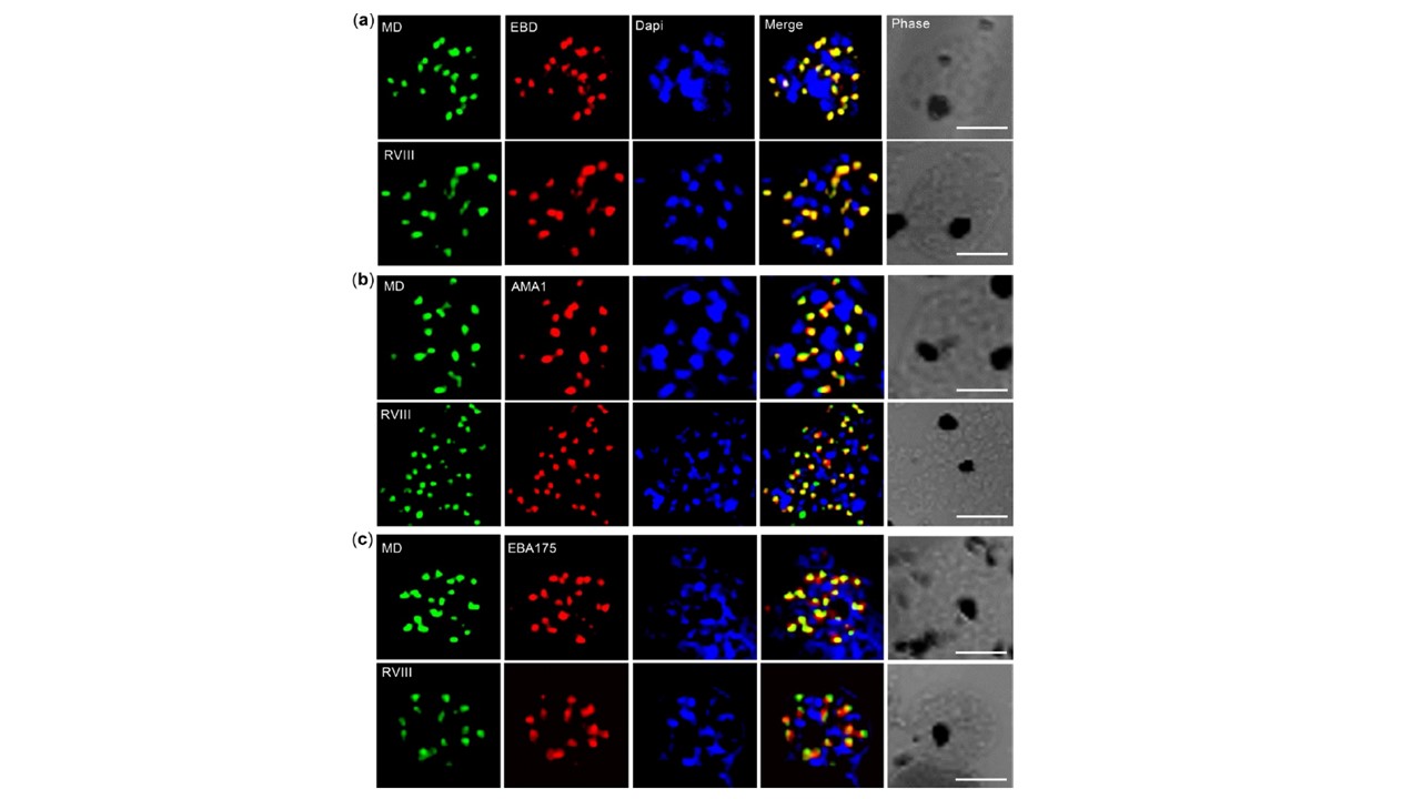
PfRH1 is localized in the rhotry in schizonts by confocal microscopy. The
co-staining was carried out in the late schizont stage W2mef by using mAbs against PfRH1 (EBD, MD and RVIII), AMA1 and EBA175. The co-staining combinations include PfRH1 mAbs EBD (a), or AMA1 (b) or EBA175 (c) in red with either MD (top panel), or RVIII (bottom panel) in green, respectively. Dapi (blue) is used as nuclear stain. Individual staining and merged images are shown for each of the mAbs. Phase. This images are shown. In the merged images, areas of overlap between the red and the green signals are shown in yellow. Scale bars = 5μm. In late stage schizonts both MD and RVIII Abs completely co-localize with EBD Ab at the apical end of the merozoite (a) with both MD and RVIII either partially or not colocalizing with micronemal proteins AMA1 and EBA175.
Gunalan K, Gao X, Yap SSL, Lai SK, Ravasio A, Ganesan S, Li HY, Preiser PR. A processing product of the Plasmodium falciparum reticulocyte binding protein RH1 shows a close association with AMA1 during junction formation. Cell Microbiol. 2020 May 25:e13232.
Other associated proteins
| PFID | Formal Annotation |
|---|---|
| PF3D7_0731500 | erythrocyte binding antigen-175 |
| PF3D7_1133400 | apical membrane antigen 1 |