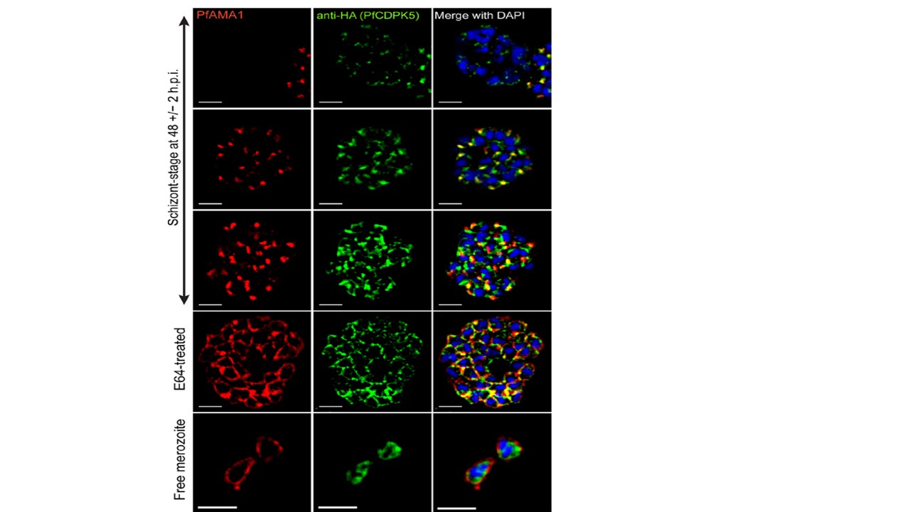
Characterization of 3D7-PfCDPK5DDKnL parasites and PfCDPK5 localization. Schizonts from [+] Shld-1 3D7-PfCDPK5DDKnL parasites were fixed, probed with anti-HA and anti-PfAMA1 antibodies, and visualized by SR-SIM. Bars, 2 mm. Initially were detected PfAMA1 expression in the micronemes and after a poorly characterized “egress trigger,” observed PfAMA1 translocation to the plasma membrane of the fully mature daughter merozoites. In 3D7-PfCDPK5DDKnL parasites with undetected PfAMA1 expression, we observed punctate cytoplasmic localization of PfCDPK5 (top row). In parasites with detected micronemal PfAMA1 localization, we observed two different staining patterns for PfCDPK5. The first was observed in parasites with weaker PfCDPK5 staining, where there was strong colocalization with micronemal PfAMA1 (second row). Given that PfCDPK5 has no known signal sequence, we hypothesize that it is located on the cytoplasmic side of these micronemes. In the second pattern, observed at higher levels of PfCDPK5 expression, PfCDPK5 retained colocalization with PfAMA1 but also exhibited additional diffuse apical staining (third row). In parasites that were beyond the “egress trigger,” PfAMA1 was translocated to the merozoite surface, and PfCDPK5 relocalized to a peripheral cytoplasmic distribution with increased signal near the plasma membrane (E64-treated row). Localization was similar in free merozoites isolated from parallel cultures grown without E64 (Free merozoite row).
Absalon S, Blomqvist K, Rudlaff RM, DeLano TJ, Pollastri MP, Dvorin JD. Calcium-Dependent Protein Kinase 5 Is Required for Release of Egress-Specific Organelles in Plasmodium falciparum. mBio. 2018 Feb 27;9(1):e00130-18.
Other associated proteins
| PFID | Formal Annotation |
|---|---|
| PF3D7_1337800 | calcium-dependent protein kinase 5, CDPK5 |