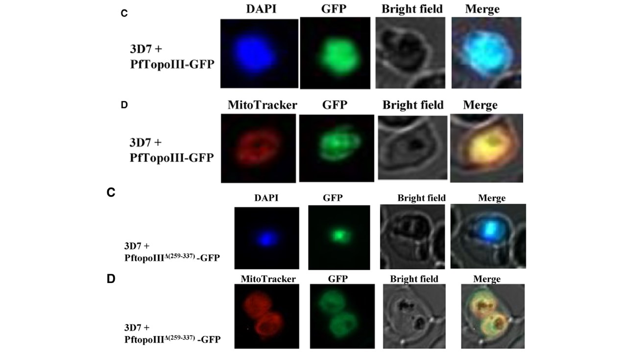
Upper panel: Subcellular localization of PfTopoIII. (C and D) Fluorescence microscopy shows the expression of PfTopoIII-GFP at the late schizont stage. Parasite nucleus was stained with DAPI (blue) while parasite mitochondria were stained with MitoTracker Red (red). Lower panel: PftopoIII(Δ259–337) shows poor association with mtDNA. (C and D) Fluorescence microscopic images show the localization of PftopoIII(Δ259–337)-GFP. Parasite nucleus was stained with DAPI (blue) and parasite mitochondria were stained with MitoTracker Red (red).
Bansod S, Bung N, Singh P, Suthram N, Choudhury H, Roy A, Bulusu G, Bhattacharyya S. Elucidation of an essential function of the unique charged domain of Plasmodium topoisomerase III. Biochem J. 2020 Nov 26:BCJ20200318.