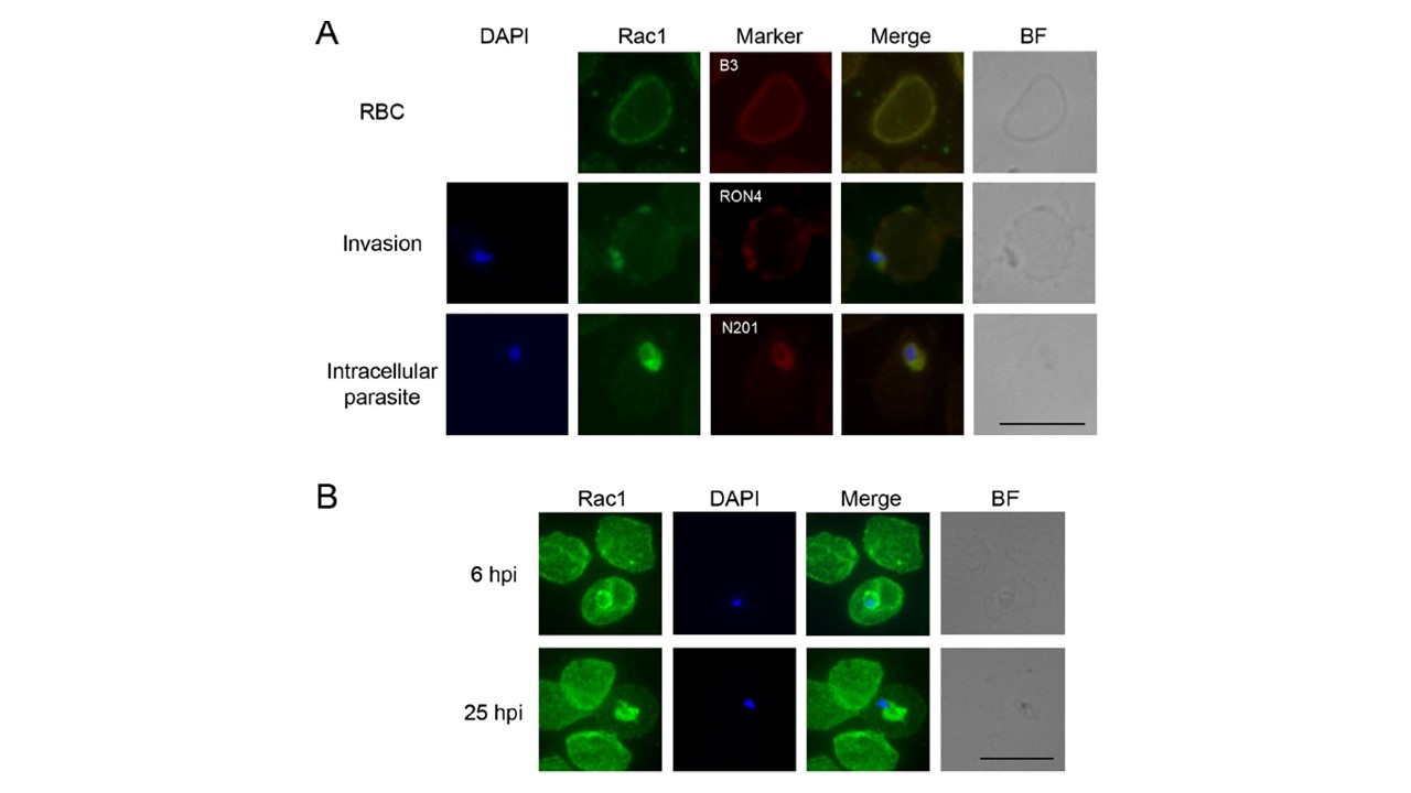
Rac1 subcellular localization. (A) IFAs of infected and non-infected human erythrocytes. Anti-Rac1 polyclonal antibody (ab#4) was used together with a primary antibody against Band3 (B3), as a marker of erythrocyte membrane, anti-RON4 antibody as a marker of the moving junction and anti-N201 as a marker of the PVM. RBC: non-infected erythrocyte. Invasion: parasite invading an erythrocyte. The nucleus has the typical bilobed shape. Intracellular parasite: parasite inside the parasitophorous vacuole. BF: Bright field. Nuclei are stained with DAPI. Different exposure times were used in each image. Scale bar: 10 μm. (B) IFA of synchronous parasites at 6 hpi and 25 hpi stained with the monoclonal anti-Rac1 R1-ab#3. BF: Bright field. Nuclei are stained with DAPI. Scale bar: 10 μm.
Paone S, D'Alessandro S, Parapini S, Celani F, Tirelli V, Pourshaban M, Olivieri A. Characterization of the erythrocyte GTPase Rac1 in relation to Plasmodium falciparum invasion. Sci Rep. 2020 10(1):22054.