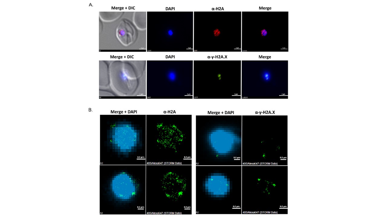
Phosphorylated PfH2A is located at distinct nuclear foci, while the nonphosphorylated PfH2A is spread throughout the nucleoplasm. Immunofluorescence of PfH2A and phosphorylated PfH2A (A; red, anti-g-H2A; green, anti-g-H2A.X; blue, DAPI; scale bar, 2mm) and superresolution STORM imaging of PfH2A and phosphorylated PfH2A (B; green, Alexa Fluor 647 staining each of the PfH2A isoforms; blue, YOYO1 staining of DNA at low resolution for orientation; scale bar, 0.5mm) in P. falciparum nuclei following X-ray irradiation (6,000 rads). The canonical PfH2A is spread throughout the nucleoplasm, its phosphorylated form is found at distinct foci (A). To further validate this observation, we performed superresolution stochastic optical reconstruction microscopy (STORM) imaging, which enabled us to image the nuclear distribution of the two forms of PfH2A in detail at the nanoscale level. This analysis clearly demonstrated the differential distribution of the two PfH2A forms in the nucleoplasm. The nonphosphorylated PfH2A is indeed spread throughout the nucleoplasm, while the phosphorylated form is much less abundant and is found at distinct nuclear foci (B).
Goyal M, Heinberg A, Mitesser V, Kandelis-Shalev S, Singh BK, Dzikowski R. Phosphorylation of the Canonical Histone H2A Marks Foci of Damaged DNA in Malaria Parasites. mSphere. 2021 6(1):e01131-20..