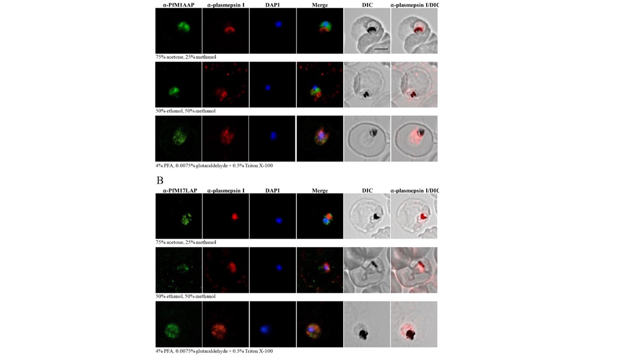
Localization of PfM1AAP and PfM17LAP in intra-erythrocytic P. falciparum 3D7 trophozoite-stage parasites. Immunofluorescence assays were carried out using air-dried blood smears fixed with 75% acetone and 25% methanol at − 20 °C for 5 min, or 50% ethanol and 50% methanol at − 20 °C for 2 min, or 4% PFA and 0.0075% glutaraldehyde for 20 min at room temperature. Fixed parasites were probed with polyclonal antibodies against (A) PfM1AAP and (B) PfM17LAP. Specific aminopeptidase staining (green, Alexa-Fluor 488) was observed in the cytosol of parasites. Parasite nuclei were visualized using DAPI (blue; 4,6-diamidino-2-phenylindole) and monoclonal antibodies against the DV marker plasmepsin I (α-plasmepsin I, red, Alexa-Fluor 594) were used as a control. Differential interference contrast (DIC) and α-plasmepsin I with DIC are shown for reference. Scale bar, 3 μm.
Mathew R, Wunderlich J, Thivierge K, Cwiklinski K, Dumont C, Tilley L, Rohrbach P, Dalton JP. Biochemical and cellular characterisation of the Plasmodium falciparum M1 alanyl aminopeptidase (PfM1AAP) and M17 leucyl aminopeptidase (PfM17LAP). Sci Rep. 2021 11(1):2854.
Other associated proteins
| PFID | Formal Annotation |
|---|---|
| PF3D7_1311800 | M1-family alanyl aminopeptidase |
| PF3D7_1407900 | plasmepsin I |