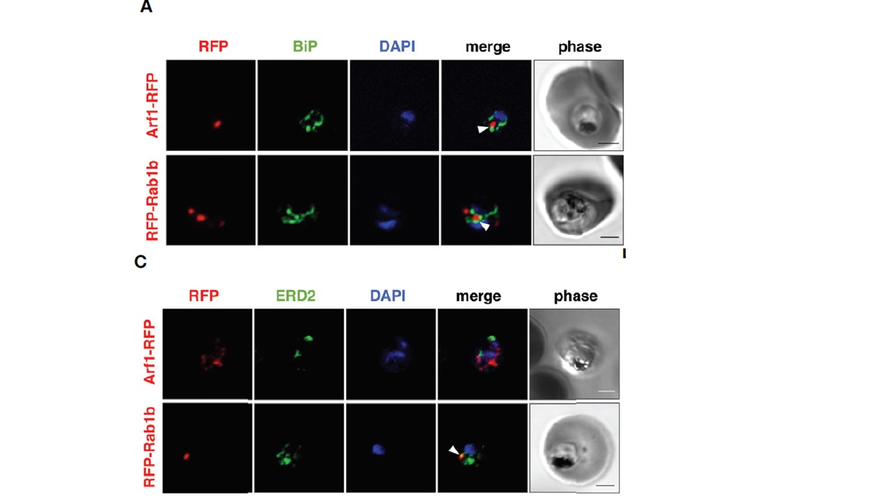
Localization of PfArf1 and PfRab1b in juxtaposition to the ER and the cis-Golgi. (A) Synchronized parasites, expressing PfArf1-RFP (upper, red) or RFPPfRab1b (lower, red), were fixed at early trophozoite stage and subjected to the indirect immunofluorescence analysis with anti-PfBiP antibody (green) and DAPI (blue). The fluorescence of PfArf1-RFP and RFP-PfRab1b is shown. PfBiP was stained with an anti-PfBiP antibody. Both PfArf1-RFP and RFP-PfRab1b localized adjacent to the PfBiP signal (arrowheads). Punctate structure signals for PfArf1-RFP and RFP-PfRab1b were closely localized with the PfBiP signals(C) Indirect immunofluorescence analysis of the localization of PfArf1-RFP (upper, red), RFPPfRab1b (lower, red), the cis-Golgi-marker PfERD2 (green), and DAPI (blue). The fluorescence from RFP-PfRab1b colocalized with the PfERD2 signal (arrowhead), but not with PfArf1-RFP. The bars indicate 2 μm. Most of the PfArf1-RFP expressing parasites did not show colocalization of the PfArf1-RFP and PfERD2 signals (32 ± 9%) (Figures 2C, D). The ratio of PfRab1b colocalization with PfERD2 was increased to 59 ± 3% in PfRab1b-RFP expressing parasites.
Taku I, Hirai T, Makiuchi T, Shinzawa N, Iwanaga S, Annoura T, Nagamune K, Nozaki T, Saito-Nakano Y. Rab5b-Associated Arf1 GTPase Regulates Export of N-Myristoylated Adenylate Kinase 2 From the Endoplasmic Reticulum in Plasmodium falciparum. Front Cell Infect Microbiol. 2021 10:610200.
Other associated proteins
| PFID | Formal Annotation |
|---|---|
| PF3D7_0917900 | PfHsp70-2 |
| PF3D7_1020900 | ADP-ribosylation factor 1 |
| PF3D7_1353600 | ER lumen protein retaining receptor |