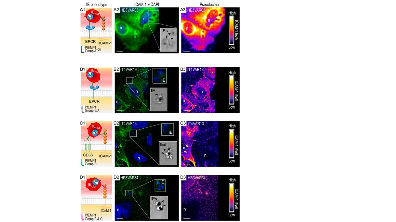
Group A+CM IEs induce clustering of ICAM-1 on brain endothelial cells. (A1–A4) IEs expressing group A+CM ICAM-1 and EPCR dual-binding PfEMP1s with a specific ICAM-1–binding motif associated with CM. IE lines in this category include HB3VAR03-IEs, 3D7 PFD1235w-IEs, and BM057-IEs. (B1–B4) IEs expressing EPCR-binding group B/ASM PfEMPs associated with SM that do not bind ICAM-1. IE lines in this category include IT4VAR19-IEs and IT4VAR20-IEs. (C1–C4) IEs expressing dual group BUM ICAM-1– and CD36-binding PfEMP1s associated with UM lacking themotif found in group A+CM PfEMP1s. IE line in this category is IT4VAR13-IEs. (D1–D4) IEs expressing group BUM or CUM PfEMP1s associated with UM that do bind ICAM-1, but lack the motif found in group A+CM PfEMP1s. IE lines in this category include HB3VAR21-IEs and HB3VAR34-IEs. See also Fig. S1 and Table 1. (A1–D1) Schematic of the four different receptor binding IE phenotypes (group A+CM, group B/ASM, group BUM, and group CUM) used in this study. (A2 and B2–D2) Wide-field (A2) and confocal images (B2–D2) of hCMEC/D3 brain microvascular cells incubated with IEs. ICAM-1 is green (FITC), and DAPI nuclei stain is blue. The bottom insets of each panel show brightfield images of IEs (black arrows) present in the framed box to the left. Top insets are an enlargement of the framed box. Blue staining in the top insets shows the presence of parasite nuclei. The images are representative of at least three independent experiments. Scale bars, 10 μm. (A2) hCMEC/D3 cells incubated with dual ICAM-1– and EPCR-binding group A+CM (HB3VAR03) IEs show ICAM-1 (green) clustered around base of the IEs. hCMEC/D3 cells incubated with (B2) EPCR-binding group B/ASM (IT4VAR19), (C2) dual ICAM-1– and CD36-binding group BUM (IT4VAR13), and (D2) ICAM-1–binding group CUM (HB3VAR34) IEs do not induce clustering of ICAM-1. (A3–D3) Pseudo-coloring of the images in A2–D2. As indicated by the color bar, areas of high ICAM-1 levels are white and yellow, while areas with low ICAM-1 levels are dark blue. Scale bars, 10 μm.
Adams Y, Olsen RW, Bengtsson A, Dalgaard N, Zdioruk M, Satpathi S, Behera PK, Sahu PK, Lawler SE, Qvortrup K, Wassmer SC, Jensen ATR. Plasmodium falciparum erythrocyte membrane protein 1 variants induce cell swelling and disrupt the blood-brain barrier in cerebral malaria. J Exp Med. 2021 218(3):e20201266.