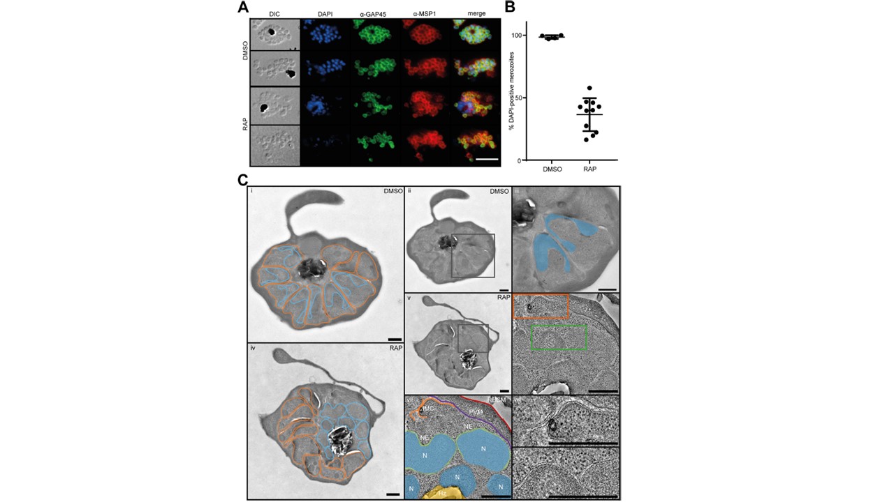
SEA1-null parasites display defective nuclear segregation during schizogony. (A) IFA images showing signals for MSP1 (a marker for the parasite plasma membrane), GAP45 (marking the IMC), and the localization of parasite DNA in mature DMSO- or RAP-treated SEA1-HA:loxP schizonts and merozoites. Scale bar, 10mm. (B) Quantification of the proportions of merozoites staining DAPI (DNA)-positive by IFA. At least 50 merozoites were counted per replicate. Totals of 554 and 682 merozoites were analyzed for the DMSO- and RAP-treated populations, respectively. Error bars, SD. (C) Electron micrographs of mature segmented DMSO- and RAP-treated SEA1-HA:loxP schizonts. (i) Example micrograph of a control (DMSO-treated) SEA1-HA:loxP schizont with merozoites outlined in orange and nuclei outlined in blue. (ii) Micrograph shown in panel i, highlighting the region shown in more detail in panel iii (black box). (iii) Region of control schizont indicated in panel ii, with C-shaped nuclei in two neighboring merozoites shaded in blue. (iv) Example micrograph of an SEA1-null schizont with merozoites and nuclei outlined as in panel i. All merozoites appear nonnucleated in this view. (v) Micrograph shown in panel iv, highlighting the region of tilt-series acquisition shown in panels vi to ix. (vi) Averages from 20 slices from the central portion of a tomogram of the region within SEA1-null schizont indicated in panel v (see also Movie S1). IMC and nuclear envelope features are present in orange and green boxes and shown in more detail in panels viii and ix, respectively. (vii) An annotated version of the tomogram highlighting the following cellular features: N (blue), nucleus; NE (green), visible nuclear envelope; Hz (yellow), hemozoin crystals in food vacuole; IMC (orange), visible IMC underlying the cytoplasmic membrane of a partially formed merozoite; PVM (purple), parasitophorous vacuole membrane; and RBCM (red), host RBC membrane. (viii) Region from the tomogram in panel vi showing more detail of the IMC formed in the SEA1-null schizont. (ix) Region from the tomogram in panel vi showing more detail of nuclear envelopes surrounding the SEA1-null nuclei that have not segregated into merozoites. Scale bars, 500 nm.
Perrin AJ, Bisson C, Faull PA, Renshaw MJ, Lees RA, Fleck RA, Saibil HR, Snijders AP, Baker DA, Blackman MJ. Malaria Parasite Schizont Egress Antigen-1 Plays an Essential Role in Nuclear Segregation during Schizogony. mBio. 2021 12(2):e03377-20.
Other associated proteins
| PFID | Formal Annotation |
|---|---|
| PF3D7_0930300 | merozoite surface protein 1 |
| PF3D7_1222700 | glideosome-associated protein 45 |