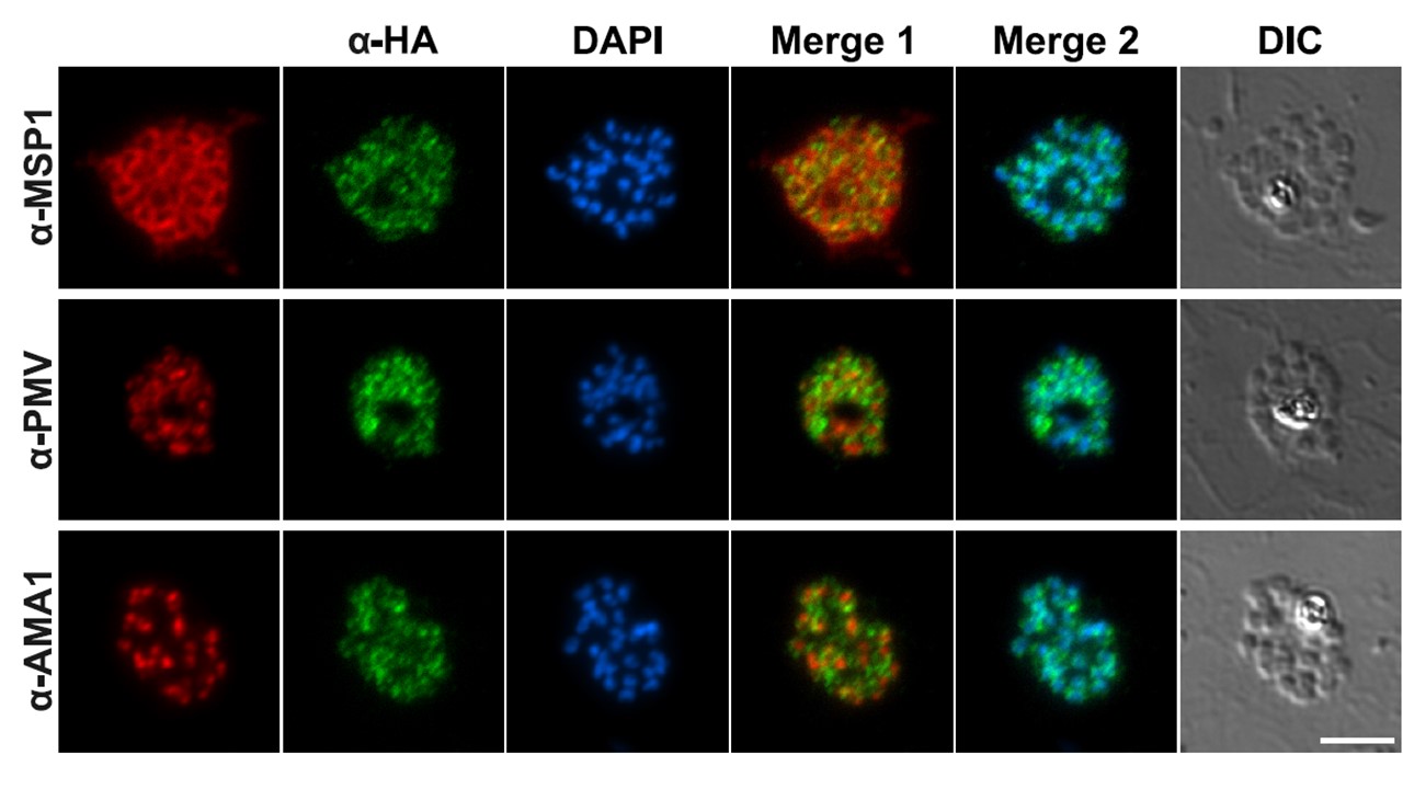
Generation of a GCa:HA-tagged line and spatiotemporal expression of GCa in P. falciparum blood stages. Dual staining IFA analysis of mature GCa:HA schizonts.
Formaldehyde-fixed thin films were stained with a-HA (green) and costained with antibodies to markers for known subcellular compartments (red): MSP1 (parasite plasma membrane, top panel), AMA1 (micronemes, middle panel), and plasmepsin V (endoplasmic reticulum, bottom panel). Scale bar, 5mm. In mature GCa:HA schizonts GCa localized to intracellular foci, but not the plasma membrane as established by costaining with a merozoite surface protein 1 (MSP1) antibody (Fig. 1F, top panel). To further characterize the nature of the intracellular compartment occupied by GCa, we costained with antibodies that react with apical membrane antigen 1 (AMA1), a micronemal marker, or plasmepsin V, an endoplasmic reticulum-resident protein. The a-HA staining showed no significant overlap with either of these markers, nor with a nuclear stain (middle and bottom panels).
Nofal SD, Patel A, Blackman MJ, Flueck C, Baker DA. Plasmodium falciparum Guanylyl Cyclase-Alpha and the Activity of Its Appended P4-ATPase Domain Are Essential for cGMP Synthesis and Blood-Stage Egress. mBio. 2021 12(1):e02694-20.