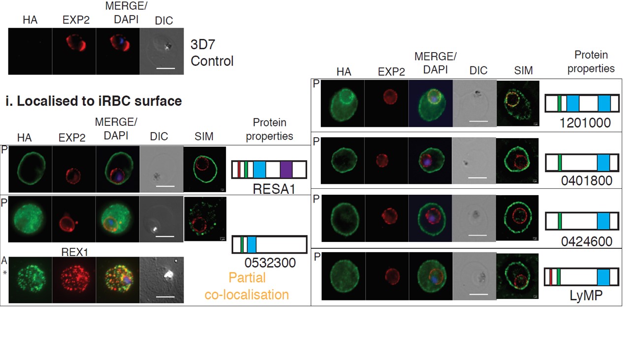
Localisation of the exported proteins. Localisation of the tagged proteins was detected via indirect immunofluorescence assays of the trophozoite stage iRBCs, when the NPP activity is greatest. All of the HA-tagged proteins were exported into the iRBC, where i) five proteins: RESA1, 0532300, 1201000, 0401800 and LyMP showed surface localisation. Structured Illumination Microscopy (SIM) analysis was used to obtain higher spatial resolution. 0532300 (*) displayed either clear surface localisation, or appeared as discrete puncta at the surface and diffused throughout the iRBC cytoplasm depending on the fixation used
Jonsdottir TK, Counihan NA, Modak JK, Kouskousis B, Sanders PR, Gabriela M, Bullen HE, Crabb BS, de Koning-Ward TF, Gilson PR. Characterisation of complexes formed by parasite proteins exported into the host cell compartment of Plasmodium falciparum infected red blood cells. Cell Microbiol. 2021 28:e13332.
Other associated proteins
| PFID | Formal Annotation |
|---|---|
| PF3D7_0401800 | Plasmodium exported protein (PHISTb), unknown function |
| PF3D7_0532300 | Plasmodium exported protein (PHISTb), unknown function |
| PF3D7_0532400 | lysine-rich membrane-associated PHISTb protein |
| PF3D7_0935900 | ring-exported protein 1 |
| PF3D7_1201000 | Plasmodium exported protein (PHISTb), unknown function |
| PF3D7_1471100 | exported protein 2 |